USF Health Byrd Alzheimer’s Center and Research Institute
Byrd Electrophysiology Core
Byrd Electrophysiology Core
The Byrd Alzheimer's Institute Electrophysiology Core is a fee-for-service facility that serves the institute and the USF neuroscience community. The Electrophysiology Core is designed to support the USF research community interested in using state-of-the-art electrophysiology techniques to investigate tissues and cells that exhibit electrical properties. It provides both standard and tailored electrophysiological services depending on the needs of the investigator. The core charges investigators for standard experimental services per animal basis. These charges are designed to pay for services, reagents, an upkeep of equipment.
Dr. Angele Parent serves as the managing faculty adviser of the Byrd Electrophysiology Core. Please contact Kassie Riley to discuss using this core facility.
Major electrophysiological applications available to the USF neuroscience community include:
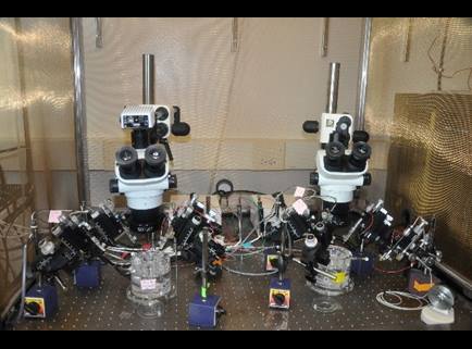
- Ex vivo field potential recording in brain slices. Long-term potentiation (LTP) and Long-term depression (LTD) in the brain, especially in hippocampus, represent useful electrophysiological correlates of learning and memory. While LTP represents long-lasting increase in signal transmission between neurons, the opposite of LTP is LTD, which produces long-lasting decrease in synaptic strength. Changes in LTP/LTD are commonly used as indicators for damage or enhancement of synaptic plasticity associated with neurodegeneration or neuroprotection. Paired pulse facilitation (PPF) is a form of short-term synaptic plasticity, which is used as a kind of presynaptic plasticity that reflects the release probability of the presynaptic neuron. PPF and input/out (IO) curves are measured to test the relationship between presynaptic and postsynaptic activity. These experiments can also be used to test the effects of drugs or chemical treatments on learning and memory.
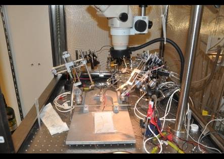
- In vivo action potential/single unit recording in anesthetized animals. The action potential conveys information from the body of the neuron to the end of the axon of the neuron. The integration and the propagation of the electrical signals (action potentials) are fundamental to nerve conduction. The action potential recording in the peripheral nerve, for example sciatic nerve, can be measured as a way of detecting changes in peripheral nerve conduction (i.e. latency, speed, amplitude, etc). Alternatively, single unit recordings can be used to study the pattern of neurons spikes in the cerebral cortex and cerebellum.
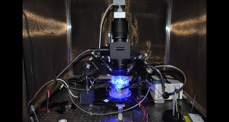
- Optical imaging in brain slices. The response to a train of electric stimulation can be imaged in ex vivo slices using Ca2+ dyes, such as Oregon green 488 BAPTA-1/AM. Ca2+ signal is the measure of the intracellular Ca2+ release. In addition, endogenous flavoprotein autofluorescence signal can be evoked and detected by a train of electric stimulation without use of exogenous dyes.The flavoprotein autofluorescence is a natural signal that comes from postsynaptic response and mitochondrial metabolism.